Why Your Calves Won’t Grow: The Fascia–Muscle Connection Most Training Programs Ignore
You train your calves consistently. You increase the weight, change the exercises, add repetitions and work both the gastrocnemius and soleus, yet they still refuse to develop the way you expect.
The conventional response is to train them harder.
At the Fascia Training Institute, we look at another variable: the mechanical environment in which the muscle is working.
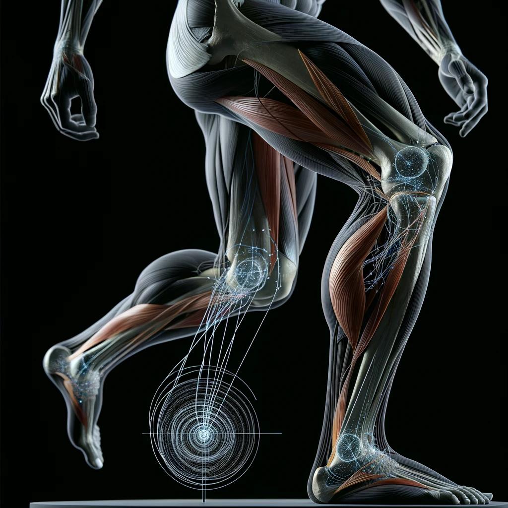
Muscle does not function independently of connective tissue. The calf operates within an integrated system involving fascia, intramuscular connective tissue, tendons, joints and neurological control. These structures influence how tissues load, lengthen, glide and transmit force.
If training is appropriate but progress remains limited, evaluating the muscle alone may be incomplete.
Your Calf Is More Than a Muscle
Traditional anatomy often teaches the body as separate structures: gastrocnemius, soleus, Achilles tendon and ankle.
That separation is useful for learning anatomy, but it does not fully represent how force moves through living tissue.
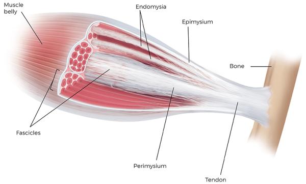
Muscle fibers exist within connective-tissue structures that include the endomysium, perimysium and epimysium. These tissues contribute to muscular architecture and force transmission.
The calf therefore cannot be understood simply as a muscle pulling on a tendon.
When the gastrocnemius and soleus contract, the muscle changes shape as it produces force. That force is transmitted through a complex interaction between contractile tissue, connective tissue, tendon and surrounding structures.
This is one reason fascia deserves greater consideration in strength, rehabilitation and performance.
Fascia Is Not Plastic Wrap
Fascia is still frequently described as wrapping around muscles.
That description is incomplete.
Connective tissue exists around and within skeletal muscle. Muscle fibers are mechanically linked with the extracellular matrix, creating pathways through which muscular force can be distributed.
Research into myofascial force transmission has challenged the traditional assumption that muscles always function as completely independent mechanical units.
This does not mean every fascial restriction causes weakness or prevents muscular development. It means muscle mechanics cannot be fully separated from the connective-tissue environment in which they occur.
What Happens When the Calf Contracts?
Place your hand around your calf and rise onto your toes.
You can feel the muscle belly change shape beneath your hand. The muscle does not simply shorten from one end to the other. Its architecture changes as force is produced and transferred toward the Achilles tendon and foot.
The surrounding connective tissues must accommodate those changes.
For a practitioner, this creates a broader assessment model. Strength is important, but so are available ankle range of motion, tissue mobility, loading mechanics and the relationship between the foot, ankle and lower leg.
The relevant question becomes whether the entire system is allowing the calf to perform effectively.
The Bodybuilding Analogy
Bodybuilders provide an interesting practical example.
Before competition, some physique athletes use massage, deep-tissue techniques and practices commonly described as “fascia stripping.”
The terminology is misleading. Fascia is not literally being stripped away, and this practice does not provide evidence that fascial treatment creates muscle hypertrophy.
What makes the example useful is the attention bodybuilders place on tissue presentation, mobility and muscular definition before stepping onstage.
Think of a fitted jacket. The structure underneath may remain unchanged, but the way the material moves over that structure affects how it appears and moves.
Human fascia is far more complex than fabric. It is living, innervated and hydrated connective tissue. The analogy simply illustrates that a muscle and its surrounding tissue environment cannot be considered completely independently.
Fascial Glide Matters
Fascial layers are capable of moving relative to adjacent tissues.
Hyaluronan, commonly abbreviated HA, is one component of the extracellular environment associated with lubrication and sliding between tissue layers.
Research has investigated how changes in HA concentration, organization and viscosity may affect normal fascial gliding. This work has contributed to the concept of fascial densification, in which changes within the extracellular matrix may alter sliding between fascial layers.
This is more precise than simply describing fascia as “tight.”
A person can experience restricted movement without fascia behaving like a physically shortened rope. Changes in tissue mechanics, sliding surfaces and the surrounding extracellular environment may contribute to what is being experienced clinically as restriction.
Why This Matters for the Calf
The calf complex manages substantial mechanical demands during walking, running, jumping, climbing stairs and maintaining balance. In training, those same structures are then asked to tolerate progressively greater loads.
The gastrocnemius and soleus work in relationship with the Achilles tendon, ankle, foot and surrounding connective tissues.
If calf development or performance is not progressing as expected, training volume is only one variable worth examining.
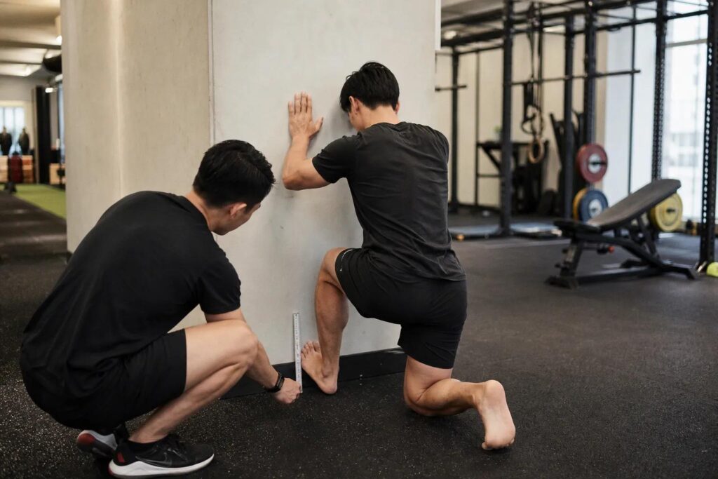
Ankle mobility may alter how a calf raise is performed. Foot mechanics can influence loading above the foot. Restrictions elsewhere in the lower extremity may change movement strategy. The person may believe they are loading the calf through a particular range while compensating around that range.
These are mechanical questions, not simply strength questions.
Can Restricted Fascia Prevent Muscle Growth?
This is where scientific precision matters.
Current evidence does not establish that “tight fascia prevents muscle growth.”
Muscular hypertrophy is influenced by mechanical loading, training volume and intensity, nutrition, protein availability, recovery, genetics, age, training history and other physiological factors.
Fascial treatment does not replace those requirements.
However, intramuscular connective tissue contributes to muscular mechanics and force transmission. The extracellular matrix also adapts to mechanical loading and participates in muscle architecture, repair and regeneration.
The more defensible question is whether altered connective-tissue mechanics, restricted range of motion or reduced tissue glide can change how a muscle is loaded during movement.
That distinction is important.
Myofascial treatment should not be marketed as a shortcut to bigger muscles. Its relevance lies in its potential relationship with movement, range of motion and the mechanical conditions under which training occurs.
Range of Motion Changes the Exercise
Consider a person performing calf raises with restricted ankle movement.
They can add weight and perform repetitions, but their available mechanics may alter how the exercise is executed.
Research examining myofascial-release interventions has reported improvements in joint range of motion. A 2024 systematic review and meta-analysis examining athletes found a moderate overall effect of myofascial-release techniques on ROM.
Other research on self-myofascial release has reported acute improvements in flexibility and range of motion without clear reductions in maximal strength or power.
These findings do not demonstrate increased hypertrophy.
They demonstrate something relevant to the FTI model: an intervention can produce a measurable change in movement.
That gives the practitioner information.
Why We Do Not Automatically Treat the Calf
A calf restriction does not automatically mean the calf is the only structure that should be assessed.
The foot, ankle, Achilles complex, lower leg, knee and hip function as mechanically related regions. Altered movement in one area can influence how load is managed elsewhere.
This does not justify claiming that every restriction in the foot causes dysfunction at the hip, or vice versa.
It does justify looking beyond the location where a limitation is reported.
The location of a symptom or restriction is useful information. It is not automatically proof of the underlying cause.
The Fascia Training Institute Approach
The FTI methodology is based on assessment and measurable change rather than assumptions about what tissue “feels tight.”
A practitioner establishes baseline movement or range of motion before an intervention. A relevant restriction is identified and an appropriate technique is applied. The movement is then reassessed.
Did range of motion change? Did the movement become easier? Did the symptom response change? Did function improve?
If the measurement does not change, that is also valuable information.
This process allows treatment to function as both an intervention and a source of clinical information.
It also reduces the tendency to repeatedly work on the same area simply because that is where the person reports tightness.
More Training Is Not Always the First Answer
When progress stalls, the fitness industry often responds by adding more load, volume, stretching or recovery techniques.
Sometimes more training is appropriate.
Sometimes the mechanics need to be evaluated first.
If someone cannot access the movement required for an exercise, adding additional resistance does not necessarily correct that limitation. It may reinforce the strategy already being used to work around it.
Addressing a measurable restriction before returning to progressive loading creates a different approach to performance.
Muscle and Fascia Belong in the Same Conversation
Progressive overload remains fundamental to muscular development. Nutrition and recovery remain essential. Genetics influence calf size and shape significantly.
None of that conflicts with fascia science.
The relevant point is that skeletal muscle is not an isolated structure. Contractile tissue and connective tissue interact mechanically, and both respond to loading.
A calf muscle is being trained within a living biological system.
When expected results are not occurring, the practitioner should be capable of evaluating more than the number of sets and repetitions.
Stop Looking at the Calf in Isolation
If your calves are not developing despite appropriate training, consider the broader mechanical picture.
Assess ankle range of motion. Examine how the foot and ankle behave under load. Determine whether the calf is moving through the intended range. Look for compensations that change how force is being distributed.
Then measure.
Intervene where indicated and measure again.
Muscular development still requires an adequate training stimulus. Fascia work does not replace that physiology.
What it can do is give practitioners another way to investigate the mechanical system in which training takes place.
The FTI Difference
At the Fascia Training Institute, practitioners are taught to move beyond isolated anatomy and investigate relationships between restriction, movement and measurable outcomes.
The objective is not to blame fascia for every problem.
It is to determine whether changing a restriction changes the result.
That distinction matters.
When movement changes after a targeted intervention, the practitioner has gained information about the system. When it does not change, the practitioner has learned something equally important.
This is how fascia education moves beyond theory and becomes clinically useful.
Find the restriction. Change the mechanics. Measure the result.
Frequently Asked Questions
Why won’t my calves grow even though I train them?
Calf development is influenced by genetics, training stimulus, exercise selection, nutrition, recovery and biomechanics. When training variables are appropriate but progress remains limited, ankle mobility, available range of motion and lower-leg mechanics may also warrant assessment.
Can tight fascia prevent muscle growth?
Current research does not establish that tight fascia directly prevents muscular hypertrophy. Fascia and intramuscular connective tissue do, however, contribute to muscular mechanics and force transmission. A restriction that changes range of motion or exercise mechanics may therefore affect how the muscle is being loaded.
Does fascia release make muscles bigger?
There is insufficient evidence that fascial release directly causes muscular hypertrophy. Research does support changes in flexibility and range of motion following some myofascial interventions. At FTI, these techniques are evaluated according to measurable changes in movement rather than claims that they directly create muscle growth.
What fascia surrounds the calf muscles?
The gastrocnemius, soleus and other structures of the lower leg interact with deep fascia and intramuscular connective tissues. The endomysium, perimysium and epimysium contribute to skeletal muscle architecture and force transmission.
Can fascia affect athletic performance?
Connective tissue contributes to muscular mechanics and force transmission. Myofascial interventions have also demonstrated effects on range of motion and flexibility in research, although outcomes depend on the intervention, population and methodology.
Should I release my calves before training?
There is no single protocol appropriate for everyone. The decision should be based on assessment. The FTI model establishes a baseline, applies an intervention where indicated and reassesses movement to determine whether the intervention produced a useful change.
Why does FTI assess more than the calf when the calf feels tight?
Because the location of restriction or symptoms does not necessarily identify the entire mechanical problem. Foot, ankle, Achilles tendon, calf, knee and hip mechanics can influence how force is distributed through the lower extremity. FTI assesses the system rather than assuming the location of the symptom is the cause.
References
Purslow PP. Muscle fascia and force transmission. Journal of Bodywork and Movement Therapies. 2010.
Turrina A, Martínez-González MA, Stecco C. The muscular force transmission system: role of the intramuscular connective tissue. Journal of Bodywork and Movement Therapies. 2013.
Wilke J, et al. Not merely a protective packing organ? A review of fascia and its force transmission capacity. Journal of Applied Physiology. 2018.
Antohe BA, et al. Effects of Myofascial Release Techniques on Joint Range of Motion of Athletes: A Systematic Review and Meta-Analysis of Randomized Controlled Trials. Sports. 2024.
Martínez-Aranda LM, et al. Effects of Self-Myofascial Release on Athletes’ Physical Performance: A Systematic Review. Journal of Functional Morphology and Kinesiology. 2024.
Professional Fascia Education
The Fascia Training Institute provides professional education for practitioners who want to understand fascia as part of an integrated mechanical and neurological system rather than treating individual muscles in isolation.
Find the restriction. Change the mechanics.