
FTI INSTITUTIONAL SCIENCE PILLAR
The Science of Fascia
Sensory Communication, Force Transmission and Adaptation
Most practitioners were trained to isolate the painful structure. Fascia demands a wider question.
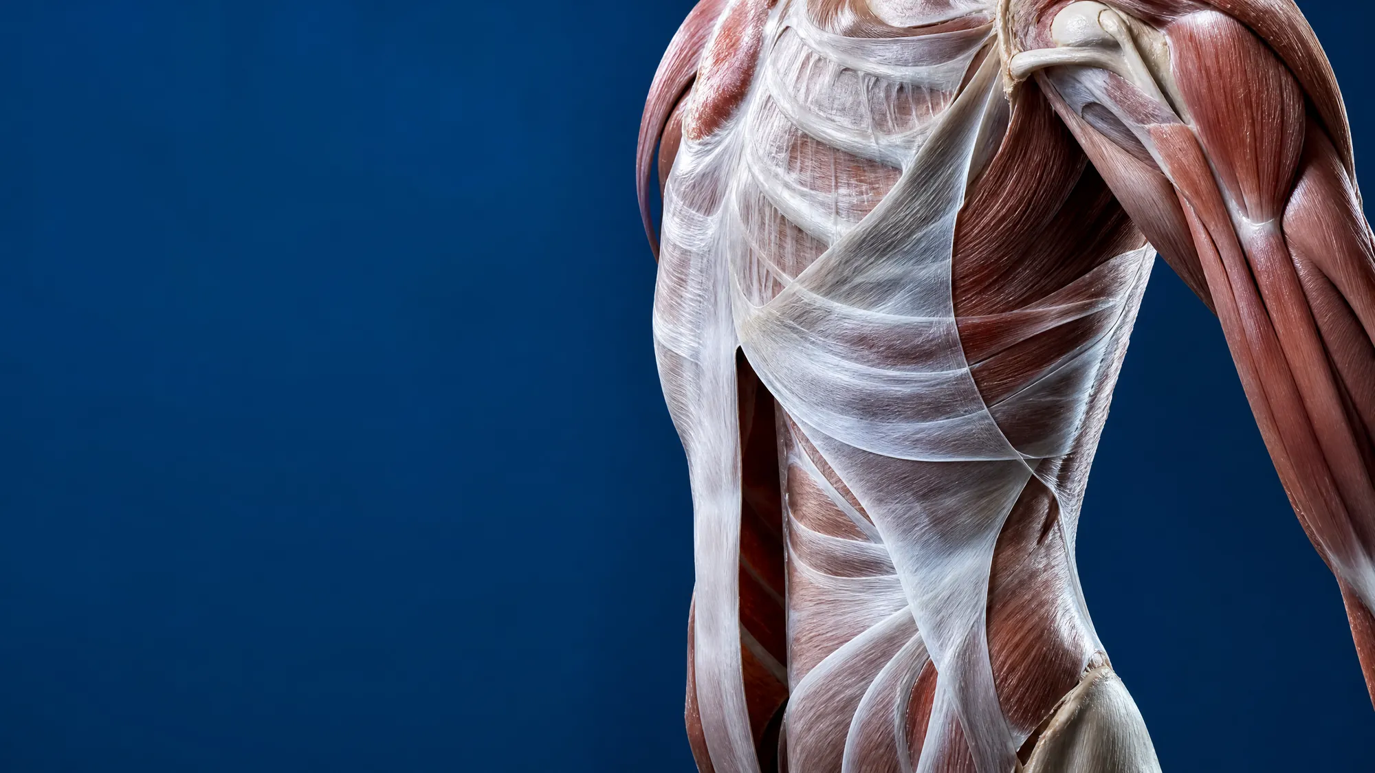
Fascia is not passive wrapping. It is living, innervated and mechanically responsive connective tissue that gives continuity to the body, supports tissue gliding, distributes load and contributes to the sensory information used to organize movement.
When fascia changes, movement can change. Force transfer can change. Sensory input can change. The location of the symptom may not reveal the location driving the limitation.

TISSUE BIOLOGY
Cells, matrix and gliding
SENSORY FUNCTION
Innervation and perception
CLINICAL REASONING
Baseline and reassessment
RESEARCH AGENDA
Signals become testable questions
DIRECT ANSWER
Fascia is a continuous connective-tissue network that surrounds, separates, supports and interconnects muscles, bones, nerves, blood vessels and organs. Its extracellular matrix, resident cells, fluid-rich interfaces and innervation allow it to respond to load, support movement and contribute to sensory signaling.
At the Fascia Training Institute, the standard is not whether a technique sounds plausible. The standard is whether the practitioner can identify a restriction, apply precise input and demonstrate a measurable change in range of motion, strength, balance, symptoms or task execution.
The Problem Is Not Always Where It Is Felt
A painful shoulder may not begin with the shoulder. A loss of cervical rotation may change after lower-body input. A muscle may test weak without being structurally weak. A client may gain range yet remain unable to control or use it.
These findings are not permission to assume that everything is fascia. They are a reason to stop treating the obvious structure without testing the wider system.
THE FTI STANDARD
If the intervention changes the system, the test should change.
If it does not, reassess the hypothesis.
Fascia Is Living Tissue—Not Inert Packaging
Older anatomy often treated fascia as material to remove in order to expose the structures considered more important. Modern research has made that view incomplete. Fascia is cellular, vascularized to varying degrees, innervated and mechanically responsive. It includes dense and loose connective tissues arranged in region-specific architectures rather than one uniform sheet.[1–3]
Its extracellular matrix contains collagen fibers that provide tensile strength, elastin that permits recoil, proteoglycans and glycosaminoglycans that influence hydration, and water-rich interfaces that support sliding between tissue layers. Fibroblasts continuously sense and respond to mechanical conditions. Through mechanotransduction, physical forces can be translated into cellular signals that influence matrix turnover and tissue organization.[3,4]
The science is no longer asking whether fascia participates in movement. The more important questions are how its different tissues behave, how local changes affect regional function, and which interventions create meaningful, durable outcomes.
Fasciacytes: Cells That Support Fascial Gliding
One important development in fascia biology is the description of fasciacytes, also called fasciocytes in general discussion. In the original 2018 study, researchers identified a fibroblast-like cell population in fascial tissue specialized in producing hyaluronan.[5]
Hyaluronan is a large, water-binding molecule found in the loose connective tissue between deep fascia and muscle and between adjacent fascial layers. It helps those layers glide. Normal movement requires more than muscle contraction and joint excursion; tissues must also slide relative to one another.
Current work is exploring how hyaluronan concentration, molecular organization, temperature, pH and mechanical load may alter viscosity and gliding behavior. The proposed calcium–hyaluronan axis is an emerging molecular framework, not yet a complete clinical mechanism.[6]

CLINICAL MEANING
A restriction may involve more than shortened muscle fibers. Altered layer-to-layer glide, local matrix behavior, protective tone and sensory input may all contribute. Test the pattern before and after an intervention rather than assuming the mechanism.
Fascia and Sensory Communication
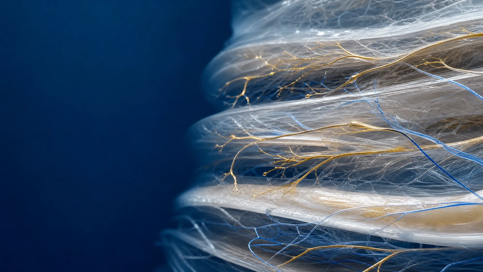
Deep fascia contains free nerve endings and other neural structures associated with nociception and proprioceptive function. Human and animal studies show that fascial tissues can be richly innervated, although density and nerve type vary by region and tissue layer.[7,8]
This does not mean fascia replaces the nervous system or communicates independently of it. It means fascia is one of the tissues from which the nervous system receives information.
- Proprioception: the brain’s estimation of body position and movement.
- Nociception: neural encoding of potentially threatening mechanical, chemical or thermal input.
- Protective motor behavior: guarding, altered recruitment and reduced movement options.
- Movement accuracy: coordination and confidence under changing load.
Pain is not a direct tissue meter. It is an output shaped by peripheral input, spinal processing, brain prediction, context and prior experience. Fascia can be one contributor to that input. It should not be treated as the sole explanation for every symptom.
Force Transmission Does Not Stop at the Tendon
Traditional biomechanics often describes muscular force traveling from muscle to tendon to bone. That pathway is fundamental, but it is not the whole picture. Anatomical and experimental studies support epimuscular and myofascial force transmission between muscles and surrounding connective tissues.[9,10]
Systematic reviews also support anatomical continuity along several proposed myofascial chains, while emphasizing that anatomical continuity does not automatically prove the magnitude or clinical importance of long-distance force transfer in every movement.[10,11]
For practitioners and performance teams, this creates a more precise working model:
- A local symptom may coexist with a restriction elsewhere in the kinetic system.
- A joint can gain range without gaining usable control.
- A muscle can test weak because force is not being organized or transferred efficiently—not only because the muscle lacks strength.
- Changing tissue input in one region can produce an immediate change in movement elsewhere. Measure it. Do not presume it.
Adaptation Under Load: Fascia Is Always Responding
Fascial tissues adapt to movement, immobilization, injury, surgery, repetitive loading, inflammation and age. Cells respond to mechanical tension and compression by changing signaling, matrix production and organization. Those responses can be useful adaptations—or become part of a persistent restriction when load exceeds recovery capacity.[3,4]
This is why “tightness” is an incomplete diagnosis. The same sensation may reflect different combinations of tissue stiffness, altered glide, protective muscle tone, sensitization, joint limitation, fatigue or neurological load.
FTI does not begin with a generic full-body sequence. It begins with the individual in front of the practitioner.
The Interstitium: Important Anatomy, Frequently Overstated
In 2018, Benias and colleagues used real-time endomicroscopy and histology to describe interconnected, fluid-containing spaces within collagen-bundle tissues beneath the skin and around organs, vessels and muscles.[12] Media reports quickly called the interstitium a “new organ.”
The more accurate interpretation is narrower and more useful. The study visualized a widespread interstitial architecture that conventional fixation may collapse. These spaces occur within connective tissues, including tissues associated with fascia. The finding does not establish that the interstitium and fascia are identical, nor did it prove a single new organ in the universally accepted anatomical sense.
It reinforced something clinically relevant: living connective tissue is hydrated, deformable and structurally different from the dry, fixed layers seen in a traditional cadaver dissection. Fluid movement, extracellular matrix behavior and tissue gliding deserve serious investigation.
What the Science Means Clinically
The research supports a systems view of movement and sensation. It does not support treating every symptom as a fascial problem or claiming that one technique can resolve every condition.
Clinically, the science supports five disciplined actions.
1. Assess
Compare position, active movement, passive range, strength, balance, symptom behavior and task performance.
2. Identify
Find the restriction that changes the system—not merely the area that feels tightest.
3. Target
Match dose, direction, pressure, position and sequence to the assessment.
4. Reassess
Repeat the same measure immediately. No change directs the next clinical question.
5. Integrate
New range without sensory control and task transfer is not a complete performance outcome.
FTI PRINCIPLE
Do not force change. Create the conditions for the system to reorganize—then measure whether it did.
Clinical Results Often Lead. Science Then Explains.
Innovation rarely begins with a finished mechanism. It begins when a repeated clinical result does not fit the existing model.
For more than three decades, FTI founder Simone Fortier has documented immediate changes in range of motion, strength, symptoms, balance, clarity and task execution after precise fascial and neurofascial input. These observations do not become irrelevant because laboratory science has not yet explained every pathway. They become research questions.
FTI’s position is direct:
- Published evidence is essential—but it is not the only place discovery begins.
- Absence of published evidence is not evidence that an observed effect is impossible.
- A repeated clinical pattern deserves structured measurement, replication and independent testing.
- A plausible mechanism is not proof of clinical efficacy.
- A clinical result should be reported at the level the data can support—no more and no less.
FTI Research: Measurable Change to Testable Questions
FTI’s research program examines outcomes at the intersection of fascia, brain function, persistent post-concussion symptoms and human performance. Its internal work includes structured case documentation, symptom inventories, pre/post functional measures and practitioner replication.
An FTI practitioner case report, Impact and Replicability of Dynamic Brain Healing Protocol Administered by Practitioners, documents outcomes after a structured protocol in a persistent-symptom case and forms part of a larger case-series program.[13] Case reports can identify signals, feasibility and questions for future study. They cannot establish causation or predict results for every person.
Next research priorities include independent replication, clearly defined comparison conditions, standardized outcome measures, longer follow-up, dose-response analysis and transparent reporting of responders and non-responders.
What Is Established—and What Is Still Being Investigated
| Evidence level | Responsible interpretation |
|---|---|
| Well supported | Fascia is connective tissue with region-specific architecture; it contributes to continuity, load distribution and tissue separation; fascial tissues contain cells, extracellular matrix and sensory innervation. |
| Supported with limits | Myofascial continuity and force transmission occur, but their magnitude and functional relevance vary. Manual and self-myofascial interventions can produce short-term changes in pain or range of motion in some populations, with heterogeneous protocols and study quality. |
| Emerging | Specialized fasciacytes contribute to hyaluronan-rich matrix. Altered matrix viscosity and glide may be clinically relevant. Molecular models such as the calcium–hyaluronan axis require further testing. |
| FTI clinical observations | Targeted fascial and neurofascial input may produce immediate changes in remote movement, symptoms, strength, balance or clarity. These signals warrant rigorous replication and mechanism studies. |
| Not established | That fascia is the cause of all disease; that the interstitium is simply another name for fascia; that one fascial restriction explains every pain condition; or that an immediate change proves a specific biological mechanism. |
Why This Matters to Human Performance
Performance is not produced by isolated muscles. It emerges from coordinated sensory input, force production, load transfer, timing, confidence and recovery capacity.
When fascial glide, sensory input or force distribution is altered, an athlete or client may still complete the task—but at a higher cost. Compensation can preserve output temporarily while reducing efficiency, precision or tolerance to repeated load.

For Practitioners
For Sport Organizations
For Clients and Athletes

FROM THE ICE
“Simone Is the Real Deal.”
After experiencing Simone Fortier’s work, former NHL player Brad May reported an immediate difference in both how his brain felt and how his body moved.
The result was compelling enough that Brad spoke directly with NHL Alumni leadership.
That recommendation led to a direct call from the NHL Alumni Association—and opened a larger conversation about Simone’s work with the brain, fascia, mobility and long-term player health.
Frequently Asked Questions
What is fascia?
Is fascia a sensory system?
What are fasciacytes or fasciocytes?
Does fascia transmit force?
Is the interstitium the same as fascia?
Can fascia cause pain?
Does myofascial release work?
What is the FTI clinical framework?
Is FTI’s work evidence-based?
Put the Science Into Practice
Choose the path that fits the work in front of you.
References
- Kumka M, Bonar J. Fascia: a morphological description and classification system based on a literature review. J Can Chiropr Assoc. 2012;56(3):179–191.
- Adstrum S, Hedley G, Schleip R, Stecco C, Yucesoy CA. Defining the fascial system. J Bodyw Mov Ther. 2017;21(1):173–177.
- Fede C, et al. A closer look at the cellular and molecular components of the deep/muscular fasciae. Int J Mol Sci. 2021;22(3):1411.
- Chiquet M, Gelman L, Lutz R, Maier S. From mechanotransduction to extracellular matrix gene expression in fibroblasts. Biochim Biophys Acta. 2009;1793(5):911–920.
- Stecco C, et al. The fasciacytes: a new cell devoted to fascial gliding regulation. Clin Anat. 2018;31(5):667–676.
- Kirkness KB, Scarlata S. Understanding Fascial Tissue on the Molecular Level—How Its Unique Properties Enable Adaptation or Dysfunction. Int J Mol Sci. 2026;27(1):160. Published online December 23, 2025.
- Mense S. Innervation of the thoracolumbar fascia. Eur J Transl Myol. 2019;29(3):8297.
- Fede C, et al. Evidence of a new hidden neural network into deep fasciae. Sci Rep. 2021;11:12623.
- Krause F, et al. Intermuscular force transmission along myofascial chains: a systematic review. J Anat. 2016;228(6):910–918.
- Wilke J, et al. What is evidence-based about myofascial chains: a systematic review. Arch Phys Med Rehabil. 2016;97(3):454–461.
- Kalichman L. Myofascial continuity: Review of anatomical and functional evidence. J Bodyw Mov Ther. 2025;45:569–575.
- Benias PC, et al. Structure and distribution of an unrecognized interstitium in human tissues. Sci Rep. 2018;8:4947.
- Fascia Training Institute. Impact and Replicability of Dynamic Brain Healing Protocol Administered by Practitioners: A Case Study. Internal case report; not peer reviewed.
- Antohe BA, et al. Effects of myofascial release techniques on joint range of motion of athletes: a systematic review and meta-analysis of randomized controlled trials. Sports. 2024;12(5):132.
- Trybulski R, et al. The effects of fascial manipulation on pain: a systematic review with meta-analysis. J Man Manip Ther. 2026;34(1):4–15.
For professional education. FTI advances research-informed education, clinically led inquiry and measurable reassessment. Clinical application remains within the practitioner’s legal scope, training and professional standards.
Last reviewed: September 2026. References are reviewed at least annually.