FASCIA • NEUROSCIENCE • BRAIN RECOVERY
Fascia, Pain and the Brain: The Missing System in Treatment and Concussion Recovery
Pain is rarely as simple as the place that hurts.
A headache may involve the cervical spine. Elbow pain may be influenced by the neck, shoulder or fascial connections above and below the joint. Persistent symptoms after a concussion may involve more than the original impact. They may include interacting changes in the brain, cervical region, vestibular and visual systems, autonomic regulation, sleep, exertion tolerance and pain processing.
This is why treating the symptom alone often produces incomplete results.
Fascia is not inert wrapping. It is a continuous, innervated connective-tissue system that surrounds, separates and connects muscles, nerves, blood vessels and organs. Around the brain and spinal cord, connective-tissue membranes interface with cerebrospinal fluid, vascular structures and lymphatic pathways.
These systems are anatomically connected. They are not clinically interchangeable.
At the Fascia Training Institute, this distinction matters. Our work does not begin with the assumption that more pressure creates more change. It begins by identifying where the system is restricted, how much neurological load the person can tolerate and what must change first.
Pain is an output. Restriction, load and impaired system regulation may be part of what is driving it.
Developed by Simone Fortier, Founder of the Fascia Training Institute and creator of Dynamic Brain Healing™.
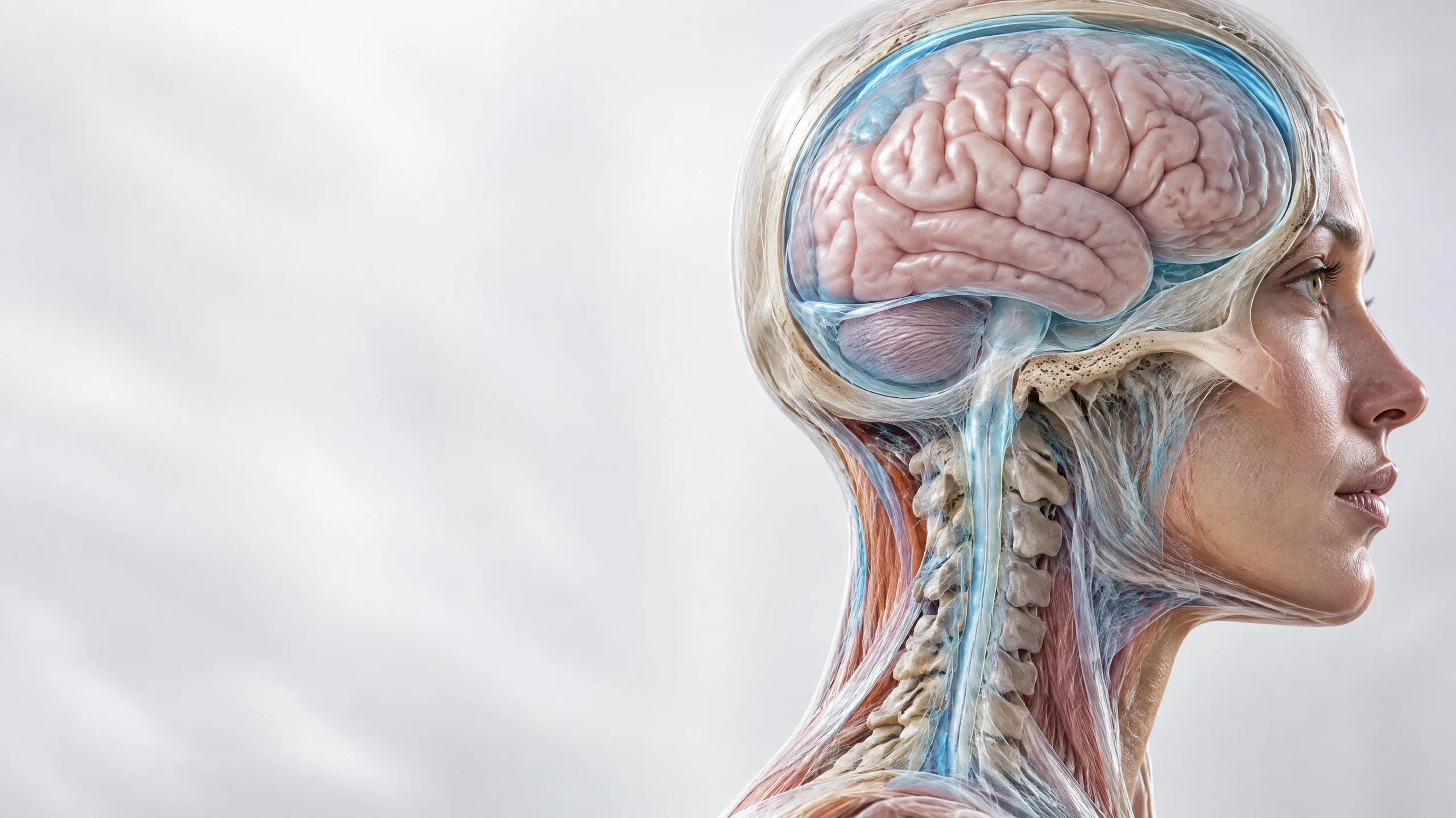
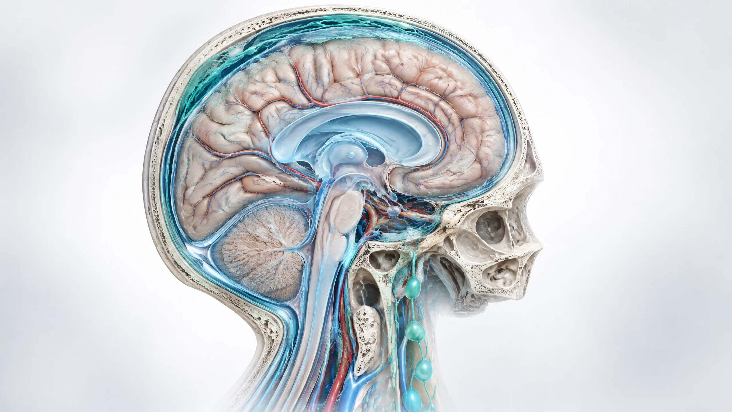
Anatomical relationship between the brain, cranial membranes, cerebrospinal fluid and upper cervical fascia.
The short answer
Fascia can contribute to pain because it is richly innervated and responsive to mechanical, inflammatory and neurological input. The connective tissues of the upper cervical region also have anatomical relationships with the spinal dura through structures known as myodural bridges. Cerebrospinal fluid and meningeal lymphatic pathways are important to brain physiology, and animal and imaging research indicates that traumatic brain injury can disturb brain-fluid transport and drainage.
What has not been established is that a manual technique can physically “flush toxins” from the brain, directly increase cerebrospinal-fluid flow or treat a concussion by itself.
The responsible clinical question is not, “Can we force the tissue to release?”
It is, “What input can this nervous system receive, integrate and use?”
Pain is an output. Restriction, load and impaired system regulation may be part of what is driving it.
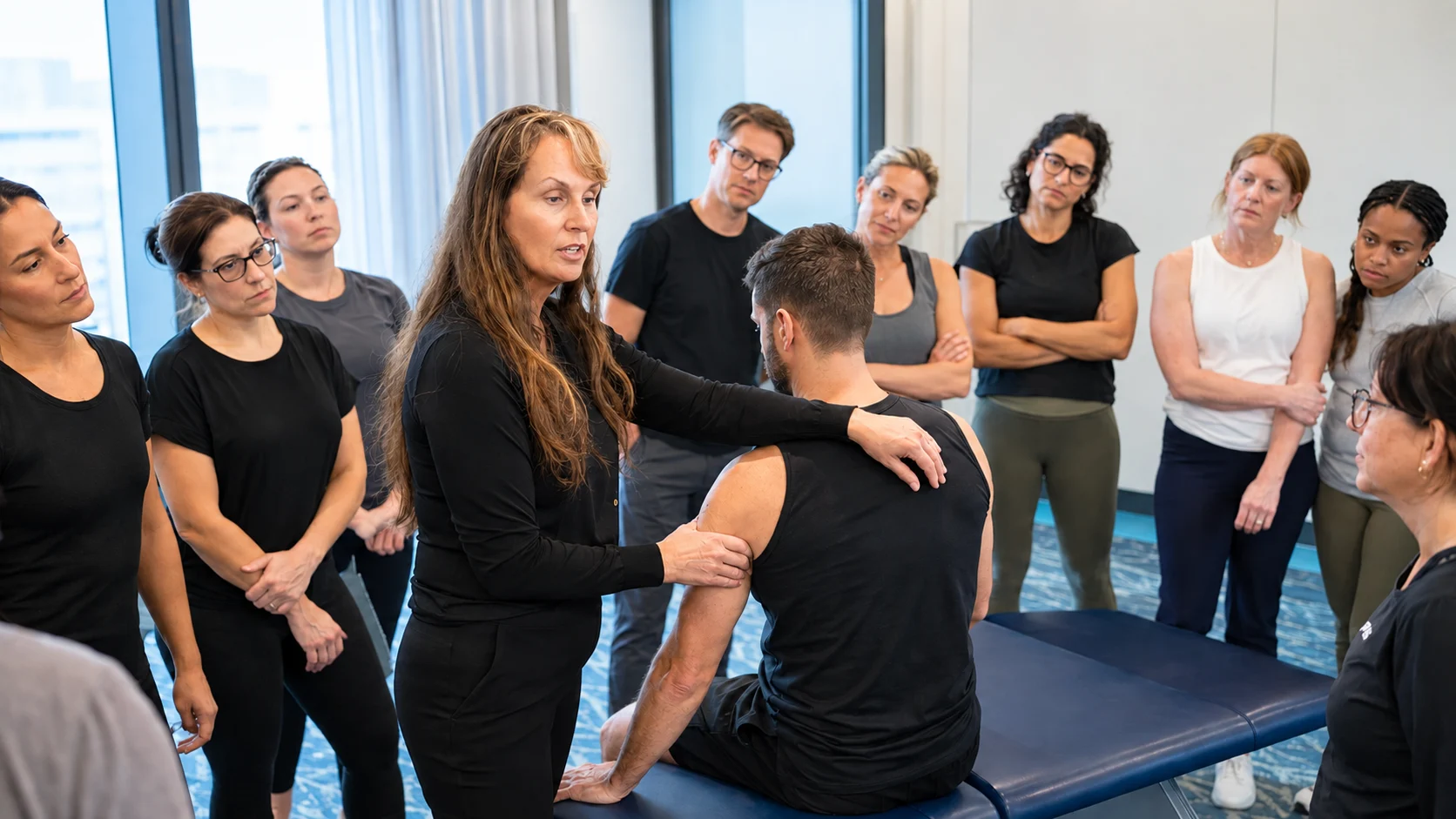
Simone Fortier leading an FTI practitioner training demonstration.
AUTHOR AND CLINICAL LEADERSHIP
Simone Fortier
Founder, Fascia Training Institute
Creator of Dynamic Brain Healing™
More than 30 years of clinical and performance experience spanning fascia, brain injury, persistent symptoms and measurable performance change. Her work has included professional and Olympic athletes, practitioners and high-performing people whose outcomes did not fit a simplified model.
Last reviewed: September 2026
What fascia actually is
Fascia is a body-wide network of connective tissue. Depending on its location, it helps:
- transmit and distribute force;
- support tissue shape and separation;
- allow layers to glide;
- house blood vessels, lymphatic vessels and sensory nerve endings;
- contribute to proprioception and interoception;
- respond to injury, inflammation, loading and movement.
Research has confirmed that deep fascia contains sensory nerve endings, including nociceptive fibers capable of contributing to pain signaling. Fascia can therefore be both a mechanical structure and a sensory interface.
That changes the treatment conversation.
When fascial layers lose their normal mobility or become sensitized, the result may involve altered movement, protective tension, reduced range of motion or pain. But fascia does not act alone. Pain is produced by the nervous system after it evaluates sensory input alongside context, previous injury, inflammation, stress, sleep, perceived threat and other biological factors.
The tissue matters. The nervous system decides what the input means.
Was fascia discovered as a “new organ”?
No. Fascia was not newly discovered in 2018.
A widely reported 2018 study used real-time imaging and specialized tissue preparation to describe previously underrecognized, fluid-filled interstitial spaces supported by collagen bundles in several tissues. The finding was important because routine slide preparation can collapse these spaces and make their living structure difficult to see.
The study expanded scientific attention to the interstitium—the fluid-containing space between and within tissues. It did not establish that fascia had suddenly become a new organ, nor that fascia causes every disease.
The more accurate conclusion is also the more useful one:
Connective tissue, interstitial fluid and lymphatic transport form a dynamic environment through which cells exchange mechanical, chemical and immune information.
This helps explain why local symptoms can be influenced by broader system conditions. It does not justify attributing every disease or every pain condition to fascia.
Fascia, pain and the nervous system
Pain is not a direct measurement of tissue damage.
It is a protective output shaped by multiple inputs. A person can have significant pain with little visible structural damage. Another person can have imaging findings without pain. After injury, the nervous system may become more protective and respond strongly to movement, pressure, light, noise, exertion or uncertainty.
Fascial restriction or sensitization may contribute by changing the quality and volume of sensory information reaching the nervous system. That may affect:
- local or referred pain;
- muscle guarding;
- movement confidence;
- joint range of motion;
- body awareness;
- tolerance for load;
- recovery after activity.
This is why the location of pain is not always the location that requires the first clinical input.
Elbow pain can be influenced by the cervical region, neural interfaces, shoulder mechanics or force transmission through the arm. Head pain can involve migraine biology, the cervical spine, jaw, visual or vestibular load, sleep, medication effects and other factors. A skilled practitioner assesses the pattern before selecting a technique.
Should fascia treatment hurt?
No. Pain is not required for effective fascia treatment.
Some people experience temporary soreness, fatigue, stiffness or a short-lived increase in symptoms after manual therapy. That reaction does not prove that toxins were released, memories left the tissue or healing accelerated. It means the system responded to the input.
The dose may have been appropriate. It may also have been too much.
Severe pain during treatment, a major loss of movement afterward or symptoms that interfere with normal function should not be celebrated as evidence of a successful release. They are signals to reassess the intensity, duration, technique, region treated and the person’s current capacity.
At the Fascia Training Institute, the aim is not to overpower restriction. It is to create the conditions in which the system can change without being driven into unnecessary protection.
FTI CLINICAL PRINCIPLE
Enough input, not maximum input
More pressure is not automatically more effective.
More pressure is not automatically more effective.
When input exceeds the nervous system’s ability to process it, guarding may increase. Pain may escalate. Range of motion may temporarily decrease. The person can leave with more neurological load than they arrived with.
History and irritability
the person’s injury and concussion history;; current pain and symptom irritability;
Cervical sensitivity and load
cervical and cranial sensitivity;; sleep, fatigue and autonomic load;
Measure, dose and integrate
range of motion before and after input;; the number of regions treated in one session;; how quickly the system responds;; recovery between sessions.
The objective is measurable change with the least unnecessary force.
Are “toxins” or memories released from fascia?
The common explanation that deep treatment releases stored toxins from fascia is not supported by current evidence.
Metabolic by-products are continuously handled through circulation, the liver, kidneys, lungs and lymphatic system. Temporary soreness after manual therapy is better described as a response to mechanical loading, nociceptive stimulation, unfamiliar input or changes in muscular and neurological activity—not as a detoxification crisis.
Likewise, memories are not known to be physically stored inside fascial tissue. Memory is encoded through nervous-system processes. However, touch, position, movement or pressure can activate sensory cues associated with a previous event. A person may experience emotion, guarding or a strong body response without the fascia itself acting as a storage vault for memory.
That distinction does not dismiss the experience.
It explains it more accurately.
What is the craniosacral system?
The brain and spinal cord are protected by three meningeal layers: the dura mater, arachnoid mater and pia mater. Cerebrospinal fluid circulates within spaces associated with these membranes and the ventricular system.
Cerebrospinal fluid helps:
- cushion the brain and spinal cord;
- maintain aspects of the chemical environment;
- transport signaling molecules and nutrients;
- participate in the movement of metabolic waste;
- interact with glymphatic and meningeal lymphatic pathways.
This is a living fluid system influenced by respiration, cardiovascular pulsation, sleep, posture and pressure gradients. The details of human glymphatic transport remain an active area of research.
It is more precise to discuss brain-fluid dynamics than to promise that a practitioner can feel or normalize a single cranial rhythm.
Where cranial fascia and the cervical spine connect
The cranium is not isolated from the neck.
Anatomical research has documented myodural bridges: connective-tissue links between suboccipital structures and the cervical spinal dura. These bridges establish a genuine structural relationship between the upper cervical soft tissues and the membranes surrounding the spinal cord.
Researchers have proposed possible roles in dural stabilization, movement-related tension and cerebrospinal-fluid dynamics. The anatomy is established. Its precise contribution to headache, dizziness and other clinical symptoms is still being investigated.
This distinction is essential.
It is reasonable to assess the cervical and suboccipital regions when a patient has headache or persistent symptoms after concussion. It is not reasonable to claim that every headache is caused by a blocked myodural bridge or that manual treatment has been proven to restore cerebrospinal-fluid circulation.
At FTI, anatomy guides assessment. Outcomes guide the next decision.
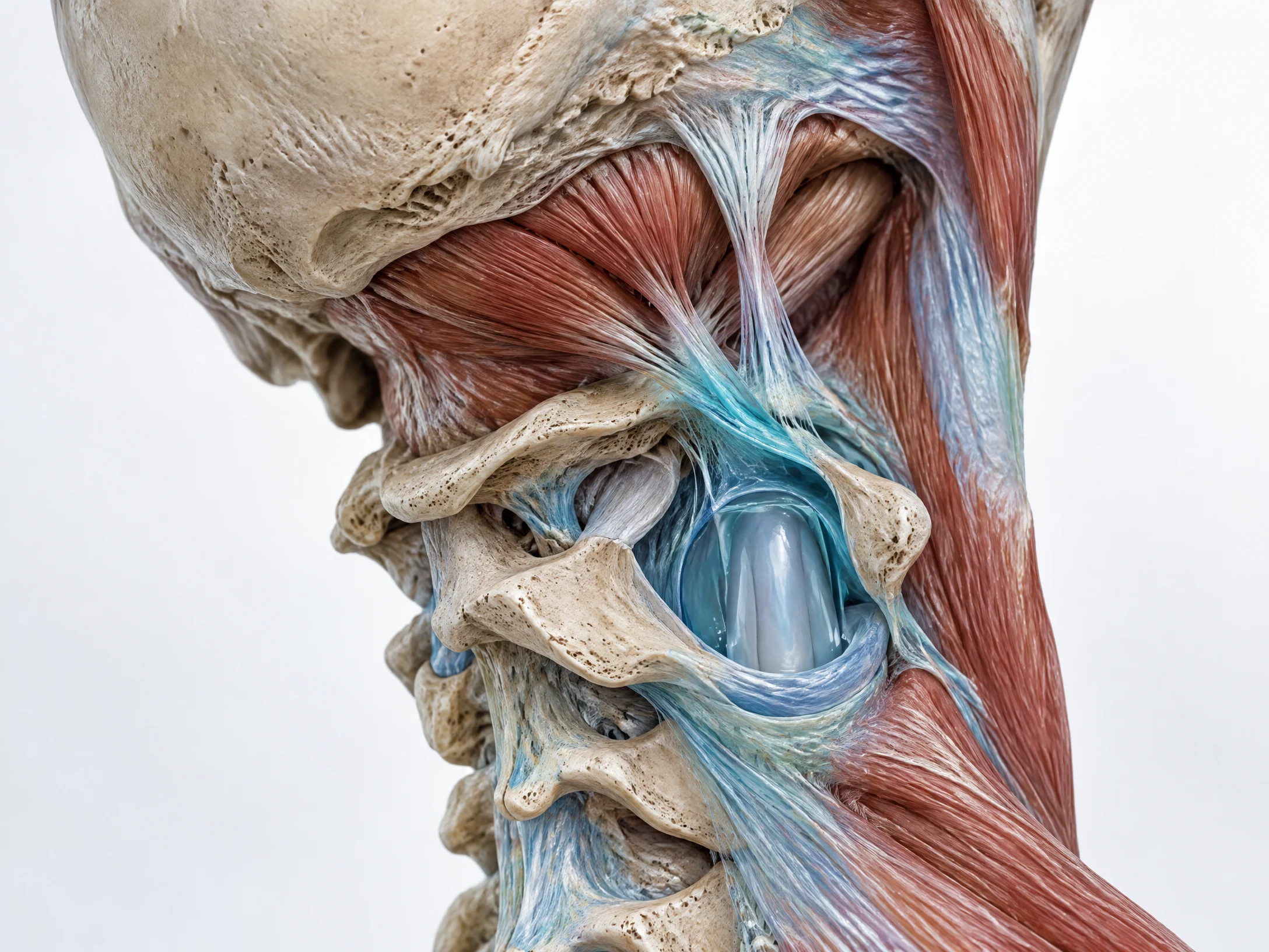
Anatomical visualization of the upper cervical soft tissues, spinal dura and myodural bridge.
Scientific visualization distinguishing cerebrospinal fluid, glymphatic exchange and meningeal lymphatic pathways.
Cerebrospinal fluid, lymphatic drainage and brain injury
Traumatic brain injury can initiate more than one event.
The primary injury occurs at the time of impact. Secondary processes may unfold afterward and can include altered metabolism, inflammation, vascular changes, ionic disruption and other cellular responses. Symptom burden can also be influenced by cervical injury, migraine, vestibular dysfunction, vision, sleep, mood and autonomic regulation.
Research has brought increasing attention to the glymphatic system and meningeal lymphatic vessels. These pathways participate in fluid exchange and waste clearance around the central nervous system. In animal studies, traumatic brain injury has impaired meningeal lymphatic drainage and worsened neuroinflammation and cognitive outcomes. Human imaging studies have also reported markers consistent with altered glymphatic function after traumatic brain injury.
This is leading-edge and clinically relevant research.
It is not yet proof that hands-on cranial or cervical techniques directly restore these pathways in humans. That question requires targeted clinical trials.
The appropriate conclusion is that brain recovery involves more than neurons alone. Connective tissue, vascular function, inflammation, sleep, cerebrospinal fluid and lymphatic pathways are part of a much larger biological system.
Concussion is a brain injury—not a single symptom
A concussion is a form of mild traumatic brain injury caused by a blow or force that disrupts brain function. Loss of consciousness is not required. Symptoms can appear immediately or evolve over hours.
Common symptoms include:
- headache or pressure in the head;
- dizziness or balance problems;
- nausea;
- light or noise sensitivity;
- blurred vision or visual strain;
- difficulty concentrating or remembering;
- mental fog or slowed processing;
- fatigue or disrupted sleep;
- irritability, anxiety or mood change;
- neck pain.
Most people improve over days to weeks, but some develop persisting symptoms that require a more detailed, individualized assessment.
The current standard is not prolonged isolation in a dark room. After an initial period of relative rest, current concussion guidance supports a gradual, symptom-limited return to daily activity and clinician-directed aerobic exercise when appropriate. Return to sport must follow a supervised progression and medical clearance.
CT and MRI are valuable when clinicians suspect bleeding, fracture or another structural injury. They do not rule out concussion, and routine imaging is not required for every uncomplicated concussion.
When a head injury needs emergency care
Seek emergency medical assessment after a head injury when there is:
- a worsening headache that does not resolve;
- repeated vomiting;
- seizure or convulsion;
- increasing confusion, agitation or unusual behaviour;
- weakness, numbness, poor coordination or slurred speech;
- one pupil larger than the other;
- loss of consciousness or increasing difficulty waking;
- fluid or blood from the nose or ears;
- significant neck pain or suspected spinal injury;
- any rapid deterioration.
Children, older adults, people taking anticoagulants and anyone with a high-force injury may require additional caution. A trained healthcare professional should direct diagnosis and acute medical management.
Why symptoms can persist after concussion
Persistent post-concussion symptoms are rarely explained by one structure or one mechanism.
Potential contributors include:
- cervical pain and altered neck function;
- vestibular or visual dysfunction;
- migraine or post-traumatic headache;
- impaired exertion tolerance;
- sleep disruption;
- autonomic dysregulation;
- anxiety, depression or post-traumatic stress;
- medication effects;
- pre-existing neurological load;
- previous head injuries;
- nutritional and metabolic factors;
- sensitized pain processing.
This is why a person may be told that a scan is normal and still be unable to think, sleep, work, exercise or tolerate light and noise normally.
Normal structural imaging does not mean the person has no injury. It means the imaging did not identify the type of structural abnormality it was designed to detect.
The Fascia Training Institute clinical framework
Simone Fortier founded the Fascia Training Institute after decades of clinical work with complex pain, performance limitations and brain injury. Her work with professional and Olympic athletes, practitioners and people who had failed to recover through standard pathways led to a consistent observation:
The system often loses capacity before it loses effort.
Dynamic Brain Healing™ was developed to assess and address the interaction between cranial and cervical fascia, neurological load, movement, symptom response and recovery capacity. It is not positioned as emergency concussion care, a replacement for diagnosis or a claim that one tissue explains every symptom.
It is a structured, brain-based and fascia-informed clinical process.
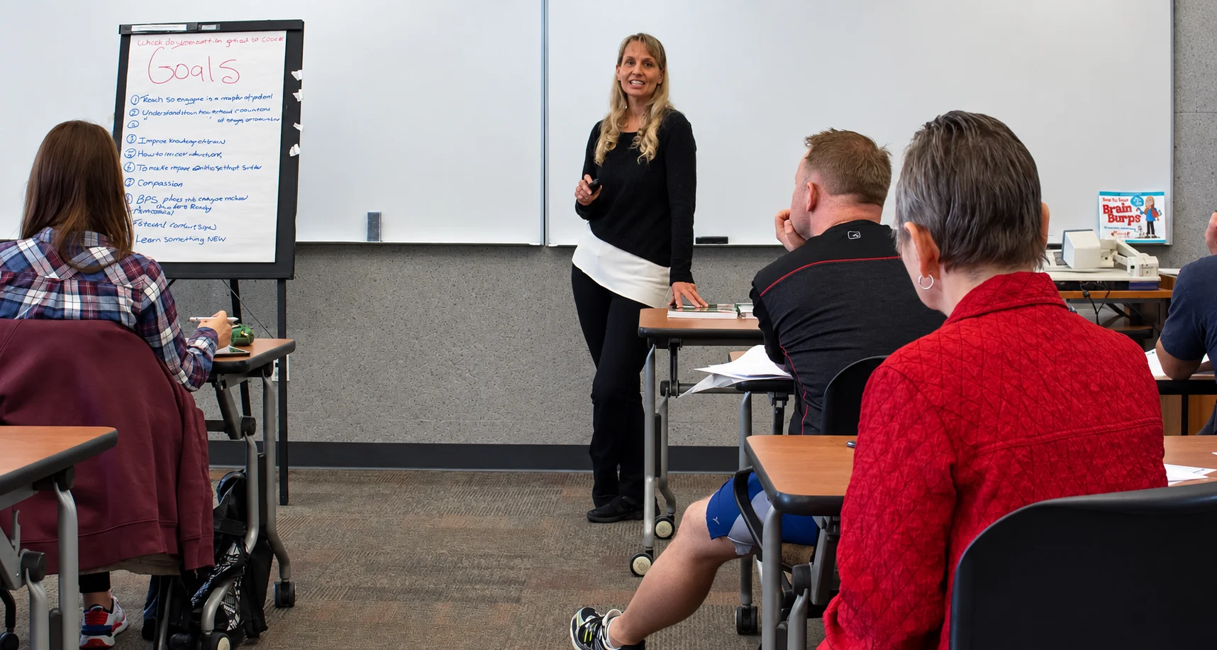
Simone Fortier teaching Dynamic Brain Healing in a Fascia Training Institute practitioner classroom.
1. Identify the dominant restrictions and symptom drivers.
01
2. Establish baseline measures, including symptoms and relevant movement.
02
3. Select the smallest effective input.
03
4. Reassess range of motion, symptoms, clarity and function.
04
5. Allow the system time to integrate before adding more load.
05
FTI clinical evidence and research direction
The Fascia Training Institute is not waiting for complex patients to fit a simplified model. It is documenting what changes when the system is assessed and treated differently.
In an FTI observational case series of 29 people with persistent post-concussion symptoms lasting longer than one year, outcomes were tracked after two Dynamic Brain Healing™ sessions. The symptom domains monitored included memory, fatigue, unrestorative sleep, brain fog, headaches, head pressure, nausea, light sensitivity and concentration. Improvements were observed across multiple tracked domains.
These findings are clinical case-series evidence, not a randomized controlled trial. They are valuable because they identify signals, measurable outcomes and the questions that should guide larger controlled studies. FTI’s research program continues to examine persistent-symptom populations, brain–fascia relationships and objective ways to measure response.
This is where clinical innovation begins: not with inflated certainty, but with a repeatable process, documented outcomes and the discipline to test what experienced practitioners observe.
OBSERVATIONAL CASE SERIES
29 cases. Persistent post-concussion symptoms for more than one year. Two Dynamic Brain Healing™ sessions. Symptoms and outcomes tracked. This is observational case-series evidence—not a randomized controlled trial.
How FTI differs from conventional craniosacral claims
The evidence for craniosacral therapy remains mixed and condition-specific. Recent systematic reviews have found methodological limitations and no dependable basis for presenting craniosacral therapy as a treatment for a long list of unrelated medical conditions.
FTI does not need inflated claims to establish authority.
Our position is more precise:
- Cranial membranes, upper cervical fascia and myodural bridges have real anatomical relationships.
- Fascia is innervated and can contribute to nociception, proprioception and movement regulation.
- Brain-fluid and meningeal-lymphatic science is rapidly developing.
- Concussion recovery is multifactorial and should be assessed as a system.
- Gentle manual input may be one component of care for selected patients.
- Clinical response must be measured rather than assumed.
- More research is required to determine how specific manual interventions affect cerebrospinal-fluid and lymphatic dynamics in humans.
That is not a smaller claim.
It is the foundation of credible clinical leadership.
What should change after treatment?
A practitioner should be able to identify what is being measured and why.
Depending on the person and the purpose of care, meaningful outcomes may include:
- improved cervical or shoulder range of motion;
- reduced headache intensity or frequency;
- better tolerance for light, movement or activity;
- reduced dizziness;
- clearer concentration;
- improved sleep;
- less guarding;
- improved capacity to work, train or complete daily tasks.
Not every change will occur immediately. Not every patient is appropriate for the same intervention. When symptoms intensify substantially or function declines, the plan must be modified and medical reassessment may be required.
Frequently asked questions
Can fascia cause headaches?
Fascial and muscular tissues in the cervical and suboccipital regions can contribute to some headache presentations, particularly when neck pain or restricted movement is involved. Headaches can also arise from migraine, concussion, medication, vascular and other causes. New, severe or unusual headaches require medical assessment.
Can neck fascia affect the brain?
The upper cervical region has connective-tissue, neural and vascular relationships with structures surrounding the brain and spinal cord. Myodural bridges provide an anatomical link to the cervical dura. The clinical effects of changing these tissues must still be evaluated symptom by symptom and patient by patient.
Does cranial treatment increase cerebrospinal-fluid flow?
Cerebrospinal-fluid movement is real and essential. Current evidence does not establish that a practitioner can reliably increase or normalize CSF flow through manual cranial treatment. This is an important area for future research, not a settled treatment claim.
Is pain necessary to release fascia?
No. Pain is not required and is not proof of an effective release. Excessive force can increase guarding and neurological load. FTI trains practitioners to use precise input and reassessment rather than intensity as the measure of quality.
Is soreness after fascia treatment normal?
Mild, short-lived soreness can occur after manual therapy. Severe pain, neurological symptoms, major loss of movement or symptoms that worsen or persist should be reported to the practitioner and medically assessed when appropriate.
Can fascia treatment cure a concussion?
No single technique should be presented as a concussion cure. Concussion requires appropriate medical evaluation and individualized rehabilitation. Fascia-informed cervical and cranial work may be considered as one part of a broader plan when clinically appropriate.
Does a normal CT or MRI mean there was no concussion?
No. Concussion is primarily a clinical diagnosis. Standard CT and MRI scans may be normal because they are used mainly to detect structural problems such as bleeding or fracture, not all functional changes associated with concussion.
What is Dynamic Brain Healing™?
Dynamic Brain Healing™ is Simone Fortier’s proprietary clinical and practitioner-training system. It integrates brain-informed assessment with precise cranial, cervical and fascial inputs, symptom tracking and measurable reassessment. It is designed for qualified practitioners and for selected clients whose symptoms or performance have not fully resolved through standard approaches.
FOR QUALIFIED PRACTITIONERS
Stop Chasing the Symptom
If your patient’s pain keeps returning, the answer may not be more pressure at the painful site.
The restriction may be elsewhere. The sequence may be wrong. The nervous system may already be overloaded. The treatment may be creating more input than the person can integrate.
Fascia Training Institute education teaches practitioners to assess the system, apply precise intervention and measure the response.
Do not force change. Create the conditions for it.
Designed for practitioners who need a more precise clinical system.
Physical therapists and physiotherapists. Chiropractors. Athletic therapists and athletic trainers. Doctors. Osteopaths. Qualified manual therapists. Eligible strength and performance professionals.
FOR PEOPLE LIVING WITH PERSISTENT SYMPTOMS
When Symptoms Persist, the System Needs a Better Assessment
If pain, headaches, dizziness, brain fog or post-concussion symptoms continue to interfere with your life, another generic treatment may not be the answer.
You may need a clearer assessment of the systems contributing to your symptoms and a plan built around your current capacity—not a protocol applied with more force.
Medical note
This page is educational and does not diagnose or treat an individual condition. Manual and fascia-based care should complement—not replace—appropriate medical assessment, emergency care, imaging when clinically indicated, or multidisciplinary concussion rehabilitation.
Research references
Benias PC, et al. Structure and Distribution of an Unrecognized Interstitium in Human Tissues. Scientific Reports. 2018;8:4947.
Fede C, et al. Fascial Innervation: A Systematic Review of the Literature. International Journal of Molecular Sciences. 2022;23(10):5674.
Sun MY, et al. Utilization of MR Imaging in Myodural Bridge Complex with Relevant Muscles: Current Status and Future Perspectives. Journal of Musculoskeletal & Neuronal Interactions. 2020;20(3):382–389.
Bolte AC, et al. Meningeal lymphatic dysfunction exacerbates traumatic brain injury pathogenesis. Nature Communications. 2020;11:4524.
Patricios JS, et al. Consensus statement on concussion in sport: the 6th International Conference on Concussion in Sport—Amsterdam, October 2022. British Journal of Sports Medicine. 2023;57:695–711.
Ceballos-Laita L, et al. Is Craniosacral Therapy Effective? A Systematic Review and Meta-Analysis. Healthcare. 2024;12(6):679.
Centers for Disease Control and Prevention. Traumatic Brain Injury and Concussion. Reviewed 2025.
FASCIA TRAINING INSTITUTE