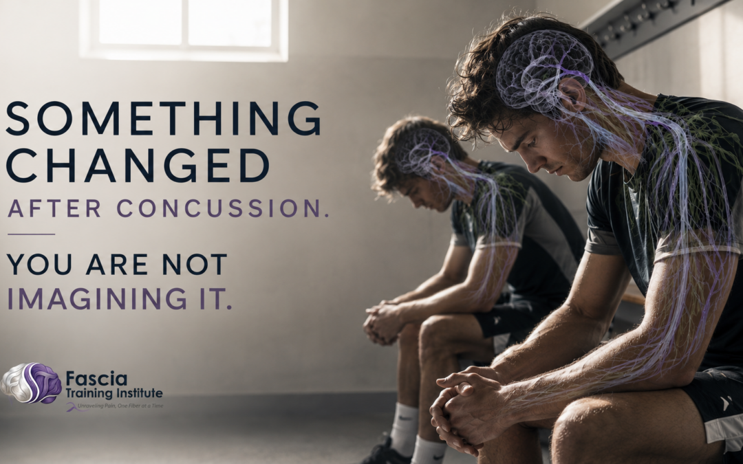
Something Changed After the Concussion. You Are Not Imagining It.
Last year I stood in front of more than 100 licensed health professionals at Lymphycon — one of the largest gatherings of lymphatic therapy practitioners in North America.
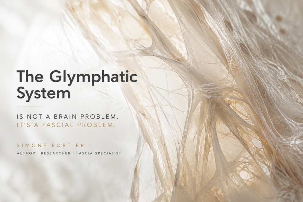
I was presenting on the glymphatic system — the brain’s primary waste clearance mechanism.
One person in the room had heard of it.
Some said it was not real.
I am sharing this not to criticize those practitioners. I am sharing it because what happened in that room reflects exactly what is happening inside the medical and rehabilitation systems managing concussion recovery right now. The practitioners are qualified. The protocols are established. And the system governing whether the brain actually recovers — the glymphatic system — is not on the assessment list.
If you are a parent whose child was cleared and is still not right. If you are an athlete who passed every test and still feels off. If you are a practitioner watching patients plateau. If you are a sporting director whose athletes are performing below baseline months after return to play.
You are not imagining it. The mechanism driving what you are observing is documented in the peer-reviewed literature. And it is not being addressed by any standard return-to-play protocol currently in use.
The symptom checklist measures above the problem. The system driving recovery is a layer beneath — one that most practitioners have never been taught to assess.
The Brain Has a Waste Disposal System. Concussion Breaks It.
The glymphatic system is a paravascular network — cerebrospinal fluid moving through channels alongside blood vessels, exchanging with interstitial fluid, and clearing the metabolic debris that accumulates in neural tissue during waking hours. Tau protein. Amyloid-beta. Inflammatory byproducts. The waste that, when it accumulates over time, is directly implicated in neurodegeneration.
It is most active during slow-wave sleep, when the brain’s extracellular space expands by up to 60%, creating the mechanical conditions under which clearance peaks. This is not a passive process. It is driven by aquaporin-4 water channels on astrocytic end-feet — molecular pumps that move fluid through the perivascular spaces surrounding blood vessels.
After a concussion, those channels are disrupted. A 2025 Frontiers in Neuroscience review of 24 studies confirmed the pre-clinical mechanism: TBI depolarizes the aquaporin-4 channels from their positions at the astrocytic end-feet. The pump that drives glymphatic clearance loses its mechanism of operation. Waste accumulates. The inflammatory cascade triggered by the initial impact cannot resolve because the system designed to clear it is not functioning at capacity.
In November 2025, the Radiological Society of North America published research showing that repeated head impacts — even sub-concussive impacts — quietly break the brain’s cleanup system. The researchers found measurable glymphatic impairment in contact sport athletes before symptoms developed. Before. The damage to the clearance mechanism preceded the clinical presentation.
This is the timeline that changes everything. The glymphatic system is being compromised during the impacts. It is not recovering on the standard timeline. And the protocols used to clear athletes back to competition are measuring symptoms — not clearance capacity.
Why Sleep Disruption After Concussion Is Not a Side Effect
One of the most consistent findings in concussion research is that sleep disruption slows recovery. Research published in the Journal of Head Trauma Rehabilitation in 2024 confirmed that athletes with disrupted sleep following concussion took twice as long to recover.
This finding has been managed as a symptom. Clinicians address sleep hygiene. They recommend melatonin. They note the disruption and monitor it.
What the glymphatic research reveals is that sleep disruption after concussion is not a side effect to be managed. It is the mechanism by which the injury persists. Slow-wave sleep is the operational window for glymphatic clearance. When that window is compromised — when the athlete cannot achieve the deep-sleep architecture the clearance system requires — the brain’s waste-removal process operates below capacity night after night after night.
The fog that will not lift. The cognitive processing that slows under load. The emotional dysregulation appears weeks after the acute phase has passed. These are not mysterious. They are the outputs of a waste clearance system that has not restarted — producing the downstream neurochemical and autonomic dysregulation that characterizes persistent post-concussion syndrome.
You are not imagining it. The mechanism is there. It is just not on the assessment form.
Sleep is not a recovery variable. It is the operational window for glymphatic clearance. When that window is disrupted, the brain cannot complete the recovery it is already trying to do.
The Tissue Nobody Is Assessing — The Dura Mater
The glymphatic system does not operate in isolation. It operates within a mechanical environment — one governed by the brain’s surrounding tissue.
The dura mater — the outermost meningeal layer encasing the brain — is cranial fascia. Dense, fibrous, mechanically heterogeneous connective tissue that is continuous with the cervical connective tissue system — the same structural interface through which rotational concussive force transmits from impact to skull to brain.
When the dura holds tension after impact — and in clinical practice, it consistently does — the pressure gradients governing cerebrospinal fluid movement are altered. The perivascular channels through which glymphatic exchange occurs are operating in a mechanically restricted environment. The aquaporin-4 channels may partially recover. Sleep may be partially managed. But if the dural mechanical environment has not been restored, the glymphatic system is attempting to function inside a compressed space.
A January 2026 randomized clinical trial protocol published in Physical Therapy — the SPINEPASS trial — is now specifically targeting dura mater function and myodural bridges in persistent post-concussion headache. The researchers identified that upper cervical hypermobility following concussion triggers an autonomic nervous system response through the dural connection. They are not treating symptoms. They are targeting the mechanical tissue environment.
This is precisely the territory Dynamic Brain Healing™ has been working in for over a decade. The dura mater as a mechanical variable in concussion recovery is not a fringe concept. It is now the subject of randomised clinical trials.
And it is still not assessed by a single standard return-to-play protocol.
What the Five-Year Data Tells Parents and Athletes
In March 2026, the journal Neurology published the largest longitudinal study of concussion outcomes in college athletes conducted to date.
3,910 former college athletes. 20 sports. Nearly half are female. Followed for five years after graduation.
Athletes with three or more concussions showed measurably worse outcomes on anxiety, depression, sleep quality, psychological distress, and concussion-related symptoms five years after leaving sport. Athletes with one to two concussions also showed worse outcomes than those with no concussions.
The researchers were precise: this is an association, not proven causation. I honor that distinction — it is scientifically correct and intellectually honest.
But here is what that limitation actually reveals for every parent and athlete reading this: we do not yet have the population-level tools to prove what the tissue-level evidence already shows is happening. The glymphatic clearance mechanism is disrupted by concussion. The dural mechanical environment restricts that clearance. Neither is assessed by standard protocols. The damage accumulates during the years of sport. And it surfaces — measurably — in the brain health of young adults in their mid-twenties who are years removed from competition.
Five years later. In their twenties. With worse sleep, worse anxiety, worse psychological distress.
If your child was cleared and is still not right, this is the research that explains why. If you are an athlete who feels like something shifted after the concussion and never fully shifted back — this is the mechanism. It is not in your head. It is in the tissue that was never assessed.
The clearance system never fully restarted. The environment that would allow it to was never restored. That is not a failure of effort or resilience. It is a gap in the protocol.
What System-Level Recovery Actually Looks Like
Dynamic Brain Healing™ was developed to address what standard protocols do not reach. Not to manage symptoms. To restore the mechanical and neurological conditions under which the brain can complete the recovery it is already physiologically capable of.
Cranial-fascial release. Targeted dural decompression. Glymphatic mobilization through mechanical restoration of the CSF environment. Vagal stimulation. Autonomic neuroregulation. Brain nutrition protocols that provide the biochemical substrate for neural repair. Each component addresses a specific layer of the environment the brain requires to clear, recalibrate, and restore.
The published evidence from the DBH prospective case series — 29 participants with persistent post-concussion symptoms exceeding 12 months, all of whom had previously undergone conventional and alternative interventions without resolution:
- Memory difficulty improved in 78% of participants
- Waking unrested improved in 74% of participants
- Fatigue improved in 72% of participants
- Cognitive fog improved in 69% of participants
- Headaches improved in 66% of participants
- Head pressure improved in 64% of participants
- Dopamine increased 32.1%
- Acetylcholine increased 19.4%
- Serotonin increased 18.2%
- GABA increased 14.7%
Two treatments. Outcomes independent of practitioner experience, participant age, and sex. Not because the method is aggressive — because it is precise. It addresses the constraint directly. When the dural restriction releases, CSF dynamics normalize. When CSF dynamics normalize, glymphatic clearance resumes. When glymphatic clearance resumes, the inflammatory debris that has accumulated since the initial impact begins to clear.
The neurotransmitter system that has been operating in a dysregulated neurochemical environment begins to stabilize. Sleep architecture improves — not as a managed symptom but as a restored function. The fog lifts not because something was suppressed but because the system that was preventing clearance was finally addressed.

The Gap Is Closeable. The Protocol Exists.
Standard concussion management was designed to determine when an athlete is safe to return to competition. That is a symptom-resolution question. It is not a system-restoration question.
Those two questions are not the same. The field has been answering the first and assuming the second follows. Thirty years of clinical practice, and a growing body of peer-reviewed research, confirms it does not.
If you are a parent reading this — the protocol gap is real, it is documented, and there is a published intervention that addresses it. You are not overreacting by pursuing something beyond the standard clearance.
If you are an athlete — what you are experiencing is a system that has not been restored. It is not a mental barrier. It is not a lack of toughness. It is a mechanical and neurological environment that has not been corrected. It can be corrected.
If you are a practitioner — the dural mechanical environment and the glymphatic clearance mechanism are now in the peer-reviewed literature with sufficient clarity that they belong in your clinical framework. The SPINEPASS trial, the Frontiers reviews, the Neurology longitudinal data — the evidence base is building rapidly.
If you are a sporting director or university athletic director — the five-year outcome data from 3,910 athletes is the kind of research that changes institutional standards. The institutions that act on it first define what best practice looks like.
The brain can clear itself. It needs the mechanical environment around it to stop working against it. Restoring that environment is not a supplement to concussion management.
It is the missing foundation of it.
Research Cited
— Broglio SP et al. Neurology, March 11, 2026 — In former college athletes, more concussions associated with worse brain health. 3,910 former NCAA athletes, 20 sports, five-year post-graduation follow-up.
— Radiological Society of North America, ScienceDaily, November 28, 2025 — Repeated head impacts may quietly break the brain’s cleanup system. Glymphatic impairment documented in contact sport athletes before symptom onset.
— Miettinen P et al. Frontiers in Neuroscience, October 2025 — Glymphatic system and mild traumatic brain injury: mini review. 24 studies 2013–2025. AQP-4 channel depolarization as primary pre-clinical driver of post-mTBI glymphatic dysfunction.
— Treleaven J et al. Physical Therapy, January 2026 — SPINEPASS Randomized Clinical Trial Protocol. Physical therapy targeting autonomic and dura mater function in persistent post-concussion headache. Myodural bridges and upper cervical dural mechanics.
— Corbali & Levey. Frontiers in Neurology, February 2025 — Glymphatic system in neurological disorders. CSF-ISF exchange; 60% interstitial space expansion during slow-wave sleep; amyloid-beta and tau clearance.
— Frontiers in Neurology, July 2025 — Meningeal enhancement following traumatic brain injury. Cranial meninges as active mechanical participants in TBI.
— Howell DR et al. Journal of Head Trauma Rehabilitation, 2024 — Sleep disturbances extend concussion recovery time. Athletes with disrupted sleep took twice as long to recover.
— DBH Prospective Case Series, Fascia Training Institute, 2025 — 29 participants, PPCS exceeding 12 months. Measurable improvements across all six post-concussion symptom domains and all four neurotransmitter indices. Outcomes independent of practitioner experience, age, and sex.